The LigMaster™
The LigMaster™ system is based on the Telos GA – II/E stress device, which has a long history of safe clinical use. |
|
The LigMaster™ software, which runs on any PC or laptop with a USB port, collects data from electronic sensors that measure displacement and - in case of ankle ligaments - rotation. These data are displayed in a graph of force against strain. From this graph, the percentage tear or laxity in the ligament is computed. The validity of the method has been verified in over 1000 clinical patients with injury to the ligaments of ankles, knees, elbows and shoulders. |
|
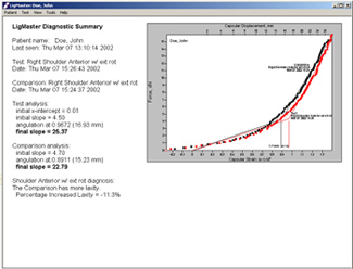 |
The extent of ligament damage is determined by comparison with the opposite, normal joint or can be compared in time series, i.e., before and after treatment to monitor ligament recovery, determine return-to-play, etc. The LigMaster™ software determines the ligament damage as percentage tear or mm laxity, using algorithms that are specific to each joint. For each joint, LigMaster calculates relevant parameters and displays them in an analysis summary. Shown here is a patient positioned in the LigMaster device for examination of the anterior glenohumeral ligaments of her right shoulder. The diagnostic box reveals that there is no clinically significant tear, but a 1.71 mm slack in her shoulder ligaments, corresponding to laxity increase of 11.3%. |
To aid in the diagnosis, affected joints are compared with the opposite, normal joint or compared over time to measure ligament recovery, or the lack thereof. Specific information on each joint can be found in the LigMaster™ user manual. |
|
LigMaster™ can perform 12 different tests to assess the ligaments most commonly injured by athletes: |
|
 |
 |
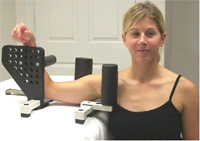
Patient positioned with the arm in 90 degrees external rotation to assess the inferior glenohumeral ligaments of the right shoulder. |
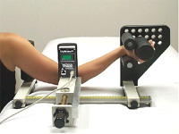
The valgus stress test is used to evaluate the medial ulnar collateral ligament of the elbow. |
 |
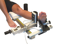 |
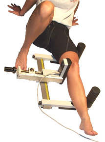
LigMaster™ can be used on the knee to evaluate the ACL, PCL, MCL and the LCL (shown). |
Patient positioned for evaluation of lateral ankle ligaments by inversion. Ankle eversion is used to evaluate the deltoid ligaments. |